


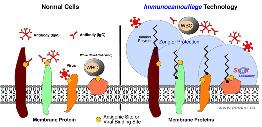
The immunocamouflage of cells and tissues is induced when selected polymers (e.g., methoxypolyethylene glycol) are "chemically glued” to proteins on the cell surface. These grafted polymers literally camouflage surface proteins and antigens as well as the inherent charge of the cell. As a result of this camouflage, receptor-ligand, cell-cell, and cell-virus interactions are blocked. However, the cell remains healthy and continues to fulfill its functions.
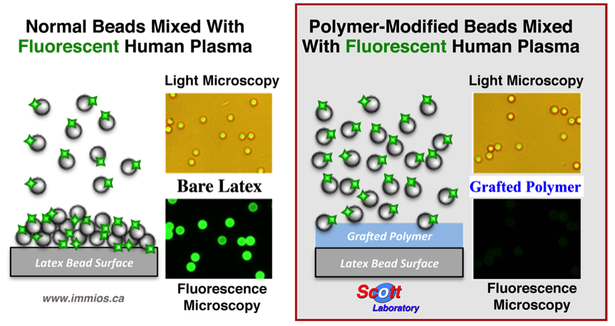
Experimentally, polymer-mediated immunocamouflage can be shown by incubating latex beads with human plasma (see below diagram). Normally, human plasma adsorbs (binds) to the latex beads, but by modifying the beads with the Immios Therapeutic polymers, the beads are camouflaged and do not bind the human plasma proteins. A similar effect occurs on the cell surface camouflaging the receptors vital for viral infection.
Immunocamouflage Mediated Immune Modulation and the Prophylactic Inhibition of Viral Infections
Human plasma will normally bind to latex beads. This is shown by the binding of fluorescently labeled plasma to normal latex beads (the spheres shown by both light and fluorescent microscopy). In contrast, the same latex beads covalently modified with polymer do not interact with the same fluorescently labeled human plasma. This camouflaging effect is highly effective and can prevent cell-cell interaction as well as viral recognition of cell receptors.
Immunocamouflage – Polymer Flexibility
Preferred Polymers
(mPEG shows the best induction of Immunocamouflage)